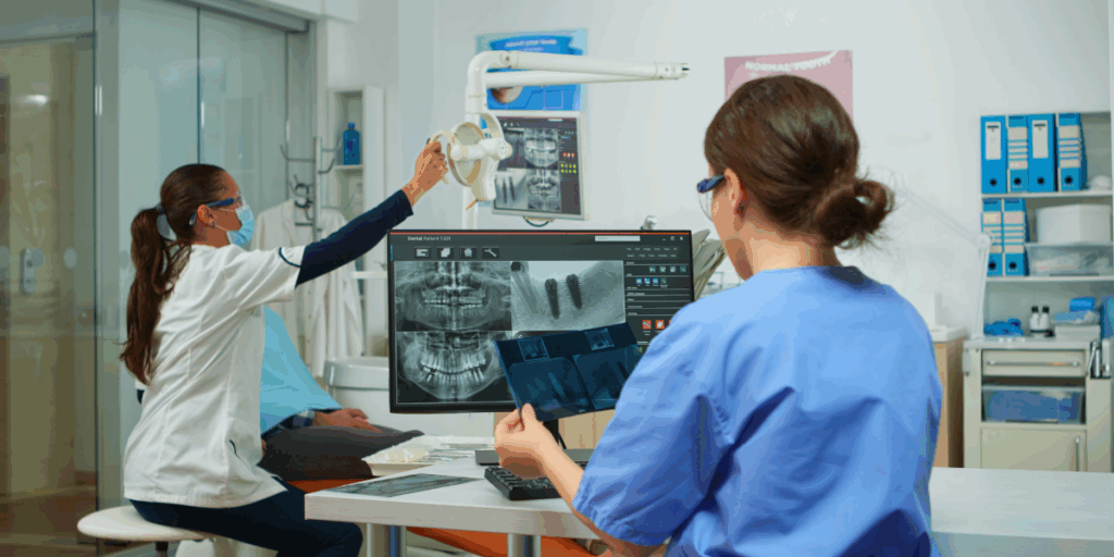
Medical imaging plays a crucial role in modern healthcare, and understanding radiological classifications can make the difference between an accurate diagnosis and missed opportunities.
With advanced medical imaging cloud storage systems now handling millions of scans daily, healthcare professionals need a solid grasp of these standardized classification systems to communicate effectively and provide optimal patient care.
What Are Radiological Classifications?
Radiological classifications serve as a universal language in medical imaging. These standardized systems help doctors, radiologists, and healthcare teams describe findings consistently across different hospitals, countries, and specialties.
Think of these classifications as detailed road maps that guide medical professionals through complex diagnostic processes. They eliminate guesswork and ensure everyone speaks the same medical language.
Major Radiological Classification Systems
BI-RADS (Breast Imaging Reporting and Data System)
The BI-RADS system revolutionized breast cancer screening by creating clear, standardized categories for mammography, ultrasound, and MRI findings.
| BI-RADS Category | Assessment | Malignancy Risk | Recommended Action |
| 0 | Incomplete | N/A | Additional imaging needed |
| 1 | Negative | <2% | Routine screening |
| 2 | Benign | <2% | Routine screening |
| 3 | Probably benign | <2% | Short-term follow-up |
| 4 | Suspicious | 2-95% | Tissue sampling |
| 5 | Highly suggestive | >95% | Tissue sampling |
| 6 | Known malignancy | N/A | Treatment planning |
Studies show that BI-RADS has reduced unnecessary biopsies by 30% while maintaining excellent cancer detection rates.
TNM Staging System
The TNM system provides a comprehensive framework for cancer staging, helping oncologists plan treatment strategies and predict outcomes.
T (Tumor): Describes the size and extent of the primary tumor N (Nodes): Indicates whether cancer has spread to nearby lymph nodes
M (Metastasis): Shows if cancer has spread to other parts of the body
RECIST Criteria (Response Evaluation Criteria in Solid Tumors)
RECIST guidelines standardize how doctors measure tumor response to treatment, making clinical trials more reliable and treatment decisions more objective.
| Response Category | Definition | Measurement Change |
| Complete Response | All tumors disappear | 100% reduction |
| Partial Response | Significant shrinkage | ≥30% reduction |
| Progressive Disease | Tumor growth | ≥20% increase |
| Stable Disease | Minimal change | Between -30% and +20% |
Specialized Classification Systems
TIRADS (Thyroid Imaging Reporting and Data System)
TIRADS helps radiologists assess thyroid nodules systematically, reducing unnecessary biopsies while catching dangerous cancers early.
Key TIRADS features include:
- Composition analysis (solid, cystic, or mixed)
- Echogenicity assessment (how sound waves bounce off tissue)
- Shape evaluation (taller-than-wide nodules raise concern)
- Margin characteristics (irregular borders suggest malignancy)
LI-RADS (Liver Imaging Reporting and Data System)
LI-RADS standardizes liver lesion reporting, particularly for hepatocellular carcinoma in high-risk patients.
Research indicates that LI-RADS implementation has improved diagnostic accuracy by 25% in liver cancer detection.
Emergency and Critical Classifications
CT Severity Index for Pancreatitis
This system grades acute pancreatitis severity using CT findings, helping emergency physicians make crucial treatment decisions quickly.
Hunt and Hess Scale for Subarachnoid Hemorrhage
Emergency radiologists use this scale to assess stroke severity and guide immediate treatment protocols.
Why These Classifications Matter
Understanding radiological classifications offers several critical advantages:
- Enhanced Communication: Standardized terminology eliminates confusion between healthcare teams across different institutions.
- Improved Patient Safety: Clear classification systems reduce diagnostic errors and ensure appropriate follow-up care.
- Better Research Outcomes: Consistent classification enables meaningful comparison of treatment results across studies.
- Legal Protection: Well-documented classification systems provide clear evidence of appropriate medical decision-making.

Staying Current with Classification Updates
Medical imaging classifications evolve constantly as new research emerges. The American College of Radiology updates major classification systems every three to five years based on the latest scientific evidence.
Healthcare professionals should regularly review updates through continuing education programs, medical journals, and professional society guidelines.
Practical Implementation Tips
Successfully implementing these classification systems requires a systematic approach:
- Create quick reference guides for commonly used classifications
- Practice applying classifications during routine case reviews
- Participate in multidisciplinary conferences to reinforce proper usage
- Stay updated through professional development opportunities
The Future of Radiological Classifications
Artificial intelligence and machine learning are transforming how we apply radiological classifications. AI systems now achieve 95% accuracy in applying BI-RADS classifications, potentially reducing human error while maintaining diagnostic excellence.
Understanding these fundamental classification systems empowers healthcare professionals to deliver superior patient care while contributing to advancing medical knowledge through consistent, reliable reporting practices.


