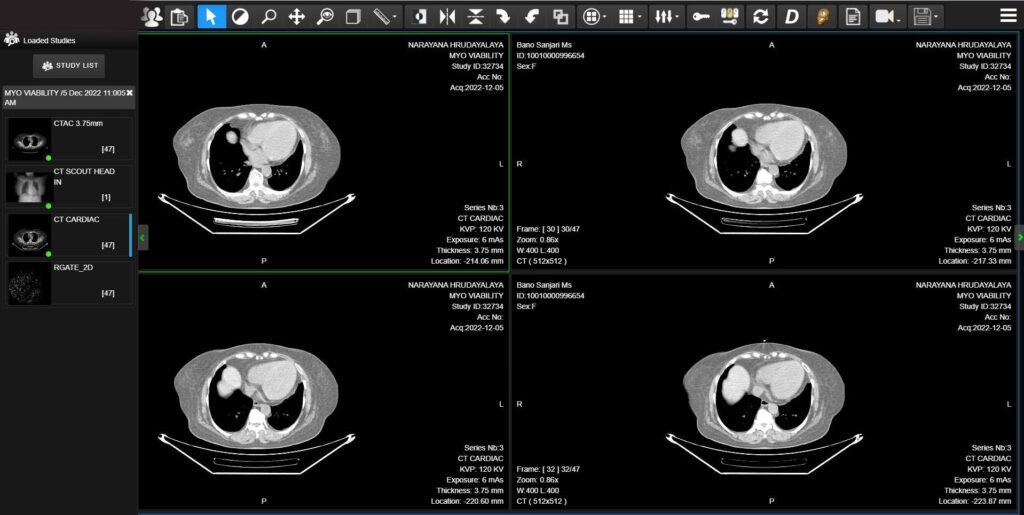
When you’re analyzing multiple medical scans side by side, every second counts.
Viewport synchronization technology in modern medical image viewer systems is transforming how radiologists compare current and prior studies, making the diagnostic process faster and more accurate than ever before.
The Hidden Problem with Traditional Image Comparison
Most radiologists don’t realize how much time they waste scrolling through images.
Research shows that radiologists can inadvertently skip clinically significant images during normal scrolling operations, especially when examining thin-slice CT scans with thousands of individual images per study.
You might think you’re seeing everything when reviewing a case, but studies reveal that traditional side-by-side comparison methods create inefficiencies that slow down diagnosis and increase the risk of missing critical findings.
How Viewport Synchronization Actually Works?
Viewport synchronization connects multiple image viewers so they move together in perfect harmony.
When you scroll through one study, all related studies automatically scroll to the corresponding anatomical location.
This spatial synchronization ensures you’re always comparing the same structures across different time points or imaging modalities.
The technology uses multi-modal rigid image registration integrated within standard PACS systems.
This means you don’t need expensive new equipment – the synchronization works with your existing setup.
Key Components That Make It Work
Image registration algorithms identify corresponding anatomical landmarks across different studies.
These algorithms account for patient positioning differences and automatically align images for accurate comparison.
Real-time tracking ensures that when you navigate to a specific slice in one study, all other synchronized viewports immediately jump to the matching location.
You can examine the exact same anatomical region across multiple time points without manual navigation.
Measurable Benefits That Impact Your Daily Work
A comprehensive study involving six radiologists reading 30 different cases revealed significant efficiency improvements when using synchronized navigation compared to traditional methods.
| Metric | Traditional Method | With Synchronization | Improvement |
| Time to read cases | Baseline | 15-25% faster | Significant reduction |
| Amount of scrolling | High | 40-60% less | Major decrease |
| Radiologist satisfaction | Moderate | High | Notable improvement |
The results were consistent across both neuroradiology and musculoskeletal cases, showing that viewport synchronization benefits multiple specialty areas. Every radiologist in the study showed measurable improvements in at least one efficiency metric.
Real Impact on Comparative Analysis Quality
Synchronized viewports eliminate the guesswork from image comparison. Instead of manually trying to find corresponding anatomy between studies, you can focus entirely on identifying changes and making diagnostic decisions.
This technology proves especially valuable when tracking disease progression over time. Oncologists using synchronized viewers can quickly assess tumor response to treatment by comparing identical anatomical locations across multiple follow-up scans.
Radiologists report feeling more confident in their diagnoses when using synchronized navigation because they can examine pathology with greater precision and spend more time on actual interpretation rather than navigation tasks.

Implementation Challenges You Should Know About
While viewport synchronization offers clear benefits, you need to consider several factors before implementation. Image registration quality depends heavily on patient positioning consistency between studies. Significant positioning differences can affect synchronization accuracy.
Training requirements are minimal for most radiologists, but your IT team needs to understand the technical requirements for proper PACS integration. Most modern systems support synchronization features, but older installations may require updates.
Network bandwidth becomes more important when synchronizing multiple high-resolution studies simultaneously. Facilities with limited connectivity might experience slower performance during peak usage periods.
The Future of Synchronized Medical Imaging
Advanced synchronization features are expanding beyond basic viewport linking.
AI-powered registration algorithms can now handle more complex anatomical variations and improve synchronization accuracy across different imaging modalities.
Multi-modality synchronization allows you to compare CT, MRI, and PET studies simultaneously, providing comprehensive views of patient anatomy and pathology.
This capability proves particularly valuable in complex cases requiring correlation between functional and structural imaging.
Recent developments include automated anomaly detection that highlights regions of change between synchronized studies, drawing your attention to areas that require closer examination.
| Technology | Current Capability | Emerging Features |
| Basic sync | Same-modality alignment | Cross-modality registration |
| Manual navigation | User-controlled scrolling | AI-assisted pathology detection |
| Static comparison | Side-by-side viewing | Dynamic change visualization |
Healthcare systems implementing viewport synchronization report improved radiologist satisfaction and reduced burnout.
When you spend less time on tedious navigation tasks, you can focus on what matters most – providing accurate diagnoses that improve patient outcomes.
The technology addresses a critical workflow challenge in modern radiology, where each CT scan can include thousands of individual images that would be nearly impossible to examine comprehensively using traditional methods.


